Ultrasound: The Gold Standard for Vascular Access
While advancing in popularity, using ultrasound imaging to support central venous catheterisation is not a new phenomenon; first described in 1978 by Ullman and Stoelting1, imaging support for accessing difficult vessels is becoming increasingly widespread. In recent years ultrasound-guided vascular access is often considered the Gold Standard as it allows medical professionals to visualise the veins and surrounding tissues in real-time. This can help to increase the accuracy and safety of the procedure, as well as reduce the risk of complications such as puncturing an artery or nerve.
Ultrasound-guided vascular access is particularly useful in patients with difficult intravenous access (DIVA), such as those with obesity, intravenous drug users, or chronic diseases. By using ultrasound, medical professionals can ensure that the catheter is inserted into the correct vein and is properly positioned, which can help to improve patient outcomes and reduce the need for repeated procedures.
What are the benefits of ultrasound-guided vascular access?
Compared with the conventional blind insertion technique, ultrasound assistance can also reduce the rates of catheterisation failure and incorrect catheter placement, increase the likelihood of success at the first pass, shorten procedure times, and reduce costs2.
According to Vezzani et al. the traditional technique relies on using anatomical landmarks rather than ultrasound guidance. Even in expert hands, it is associated with a high failure rate and a host of complications, ranging from mechanical problems (which occur in 5–19 % of cases) to infectious and thrombotic events (2–26 %)2.
Central Venous Catheterisation
Ultrasound-guidance has been promoted as a method for reducing the risk of complications during central venous catheterisation3. The central vessels that are most frequently catheterised are the internal jugular, subclavian, and femoral veins.
Internal Jugular Vein
During internal jugular venous catheterization, ultrasound-guidance reduces the number of mechanical complications, the number of catheter-placement failures, and the time required for insertion3.
Subclavian Vein
Real-time ultrasound-guided cannulation of the subclavian vein has advantages over the landmark-guided technique4,5,6. In a randomised control trial comparing the use of ultrasound-guided subclavian vein cannulation in 200 patients vs the landmark method in 201 patients, the study concluded that ultrasound-guided cannulation of the subclavian vein in critical care patients is superior to the landmark method and should be the method of choice6.
Femoral Vein
Ultrasound-guided cannulation of the femoral vein has been reported to reduce the time required for the procedure, reduce the number of passes needed to puncture the vein, and minimise complications such as arterial puncture or haematoma7.
Using ultrasound to assist with midline placement:
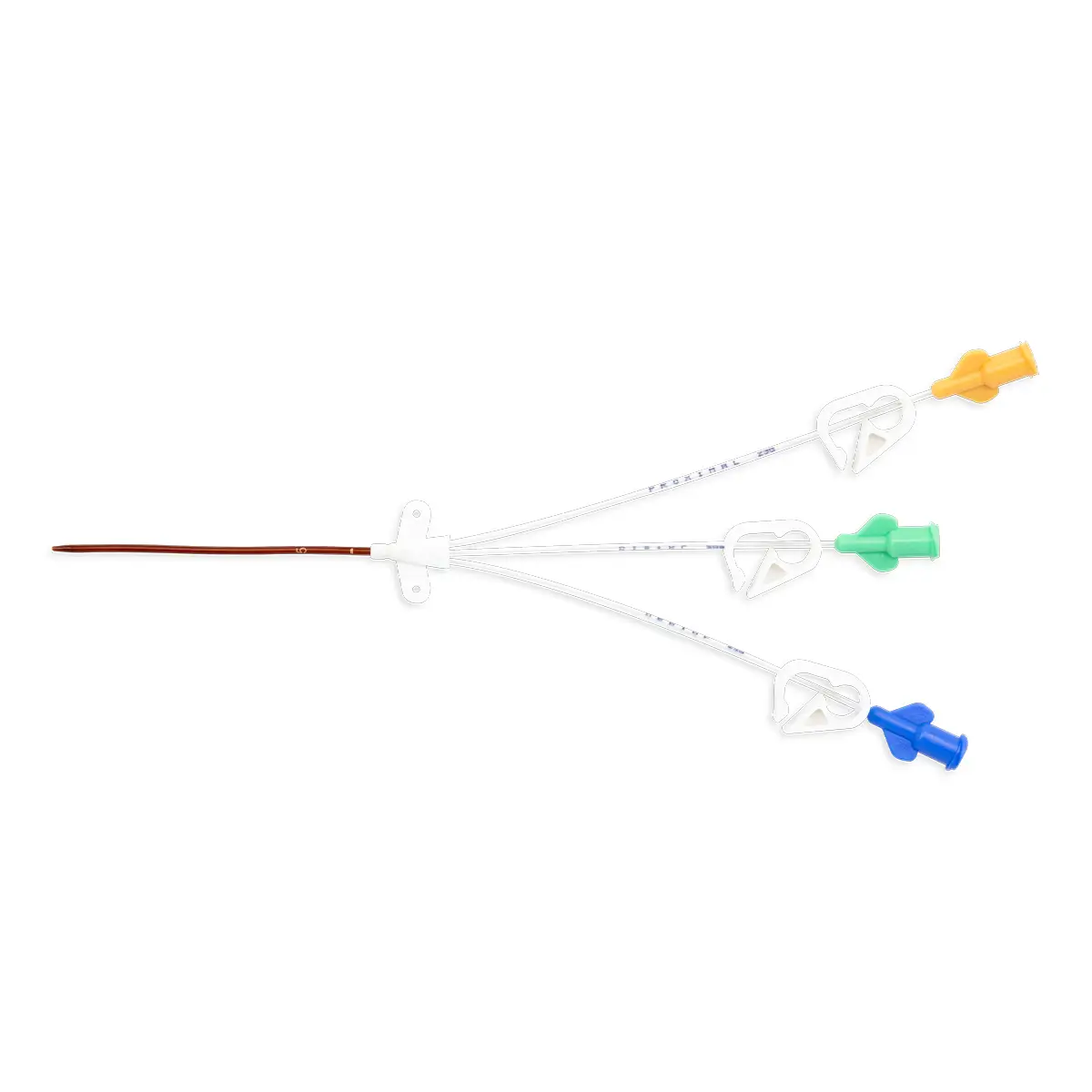
Recently, insertion of a midline catheters is recommended to be performed under ultrasound-guidance and placing it in the arm8. A study of DIVA patients in the surgical intensive care unit has shown that ultrasound-guided midline catheters are not only cost-effective, but can help facilitate early central line removal and, therefore, may reduce central line-associated bloodstream infections (CLABSI) rates9.
With proven cost savings for the NHS10, midlines are becoming increasingly popular. Using ultrasound to assist in midline placement could bring additional benefits to hospitals and Trusts.
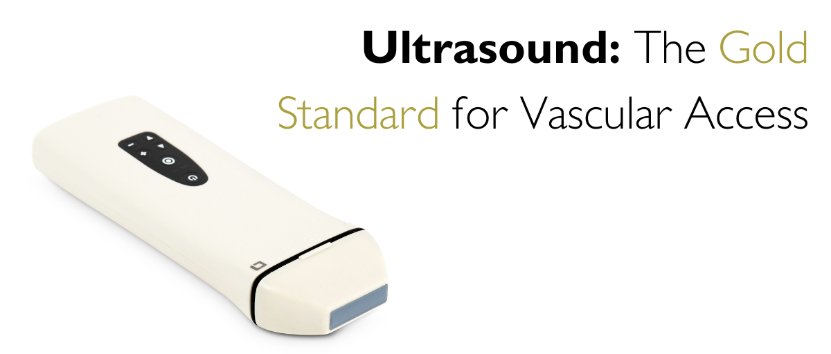
vysion Andy, the pocket ultrasound system for vascular access
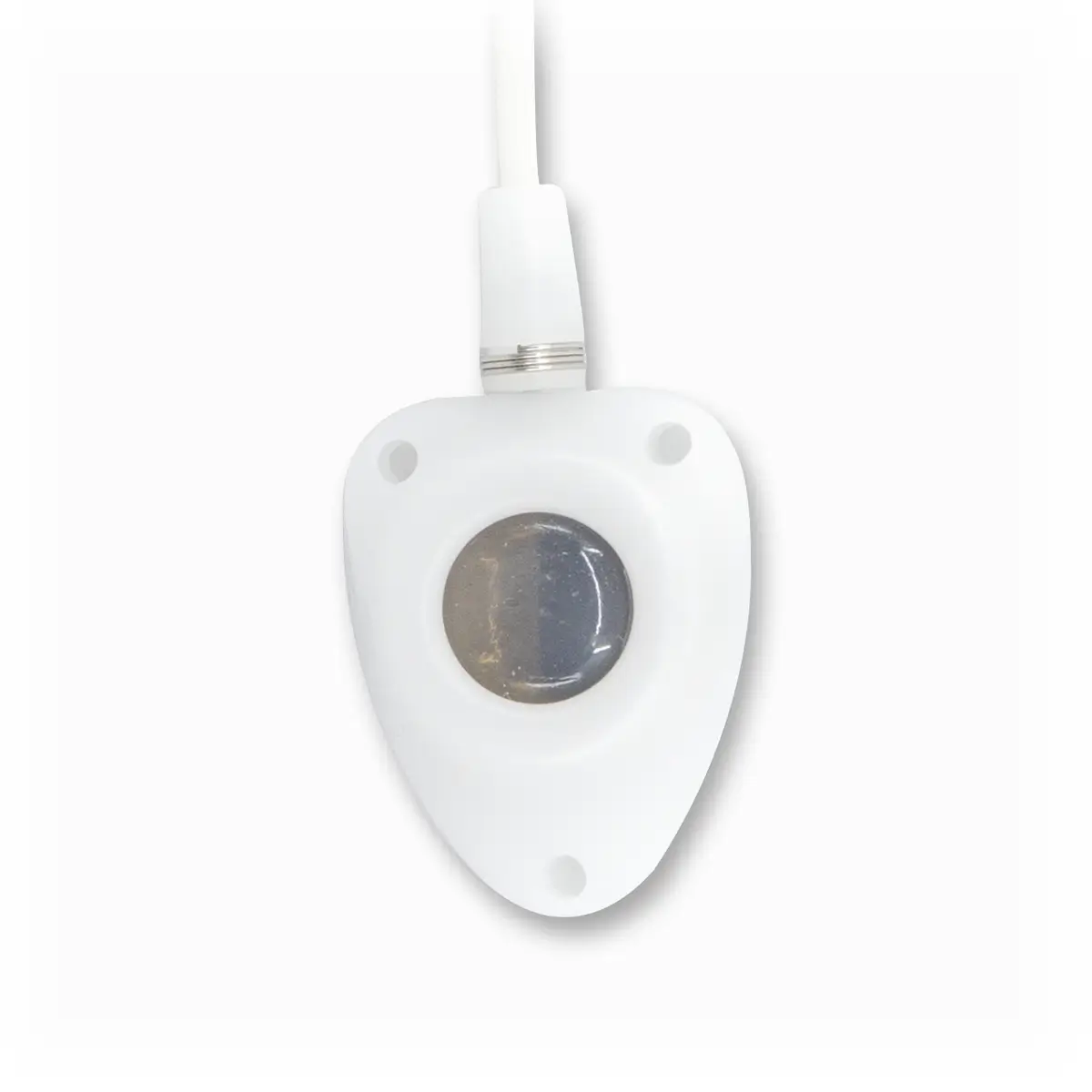
Vygon UK is excited to introduce our newest ultrasound system, vysion Andy. This lightweight pocket ultrasound supports with all vascular access procedures from echo-tracking and ultrasound-guided puncture to tip location.
Advantages of vysion Andy:
The main advantages are the benefits to the patients, including a lower risk of insertion complications (e.g. arterial puncture, nerve damage, haematoma, pneumothorax in subclavian and internal jugular) and a lower risk of catheter failure. In addition to a higher rate of first-attempt success, reducing the procedure time. Further features of vysion Andy include:
- Wifi built in, no need for an external signal
- High quality Image
- Easy mobility
- Small (170mm x 56mm x 22mm) and lightweight (only 220g)
- Compatible with PC, tablet, and smartphones
- Waterproof
- Low energy consumption with 8 hours of continuous scanning time
References
[1] Ullman JI, Stoelting RK. Internal jugular vein location with the ultrasound Doppler blood flow detector. Anesth Analg. 1978 Jan-Feb;57(1):118. doi: 10.1213/00000539-197801000-00024. PMID: 564628.
[2] Vezzani A, Manca T, Vercelli A, Braghieri A, Magnacavallo A. Ultrasonography as a guide during vascular access procedures and in the diagnosis of complications. J Ultrasound. 2013 Oct 29;16(4):161-70. doi: 10.1007/s40477-013-0046-5. PMID: 24432170; PMCID: PMC3846948.
[3] McGee DC, Gould MK. Preventing complications of central venous catheterization. N Engl J Med. 2003 Mar 20;348(12):1123-33. doi: 10.1056/NEJMra011883. PMID: 12646670.
[4] Lalu, Manoj M. MD, PhD, FRCPC1,2; Fayad, Ashraf MD, FRCPC1,2; Ahmed, Osman MD3; Bryson, Gregory L. MD, MSc, FRCPC1,2; Fergusson, Dean A. PhD2,4,5,6; Barron, Carly C. MSc3; Sullivan, Patrick MD, FRCPC1; Thompson, Calvin MD, FRCPC1 on behalf of the Canadian Perioperative Anesthesia Clinical Trials Group. Ultrasound-Guided Subclavian Vein Catheterization: A Systematic Review and Meta-Analysis. Critical Care Medicine 43(7):p 1498-1507, July 2015. | DOI: 10.1097/CCM.0000000000000973
[5] Brass P, Hellmich M, Kolodziej L, Schick G, Smith AF. Ultrasound guidance versus anatomical landmarks for subclavian or femoral vein catheterization. Cochrane Database Syst Rev. 2015 Jan 9;1(1):CD011447. doi: 10.1002/14651858.CD011447. PMID: 25575245; PMCID: PMC6516998.
[6] Fragou M, Gravvanis A, Dimitriou V, Papalois A, Kouraklis G, Karabinis A, Saranteas T, Poularas J, Papanikolaou J, Davlouros P, Labropoulos N, Karakitsos D. Real-time ultrasound-guided subclavian vein cannulation versus the landmark method in critical care patients: a prospective randomized study. Crit Care Med. 2011 Jul;39(7):1607-12. doi: 10.1097/CCM.0b013e318218a1ae. PMID: 21494105.
[7] Kwon TH, Kim YL, Cho DK. Ultrasound-guided cannulation of the femoral vein for acute haemodialysis access. Nephrol Dial Transplant. 1997 May;12(5):1009-12. doi: 10.1093/ndt/12.5.1009. PMID: 9175060.
[8] Yokota T, Tokumine J, Lefor AK, Hasegawa A, Yorozu T, Asao T. Ultrasound-guided placement of a midline catheter in a patient with extensive postburn contractures: A Case report. Medicine (Baltimore). 2019 Jan;98(3):e14208. doi: 10.1097/MD.0000000000014208. PMID: 30653177; PMCID: PMC6370112.
[9] Deutsch GB, Sathyanarayana SA, Singh N, Nicastro J. Ultrasound-guided placement of midline catheters in the surgical intensive care unit: a cost-effective proposal for timely central line removal. J Surg Res. 2014 Sep;191(1):1-5. doi: 10.1016/j.jss.2013.03.047. Epub 2013 Apr 6. PMID: 24565504.
[10] Pioneering Midline Catheter from Vygon Saves NHS Trust Over £75k. 2023 May 9th. https://vygon.co.uk/midline-catheter-gloucestershire-case-study/

